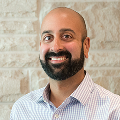
Ali Khan
Robarts Scientist, CFMM
Associate Professor, Department of Medical Biophysics and Medical Imaging, Western University

| Degree: | Ph.D. Engineering Science (Simon Fraser University ), B. Sc. Engineering Science Simon Fraser University |
| Email: | alik@robarts.ca |
| Phone: | 519.661.2111 X 24280 |
| Office: | RRI 1240A |
| Wesite | www.khanlab.ca |
Medical images play a critical role in the diagnosis and monitoring of disease, and the planning and guidance of surgical therapies. Our group develops and applies sophisticated image processing and analysis techniques to extract, quantify, and distill information from medical images, ultimately leading to more accurate diagnoses and more precise surgical interventions. We carry out multi-disciplinary research that spans across several domains, with applications in epilepsy, cancer, cardiovascular disease, and neuroscience.My research is focussed on the development of intelligent image processing and analysis technologies, and their application in the diagnosis and treatment of neurological disorders.
Research Summary
Research Questions
Can ultra-high field 7T MRI help us better understand the structure and function of the hippocampus?
The hippocampus is a brain structure that has been implicated in many different neurological conditions including epilepsy and Alzheimer’s disease, where it plays a role in both diagnosis and treatment. Emerging evidence is revealing that the different sub-structures, or subfields, of the hippocampus are uniquely affected by disease, and that complete understanding of the disorder requires careful consideration of the complex hippocampal anatomy and circuitry. My lab is investigating novel imaging and image analysis techniques using 7T MRI to characterize and quantify structure and function of the hippocampus to improve clinical care of epilepsy patients.
Can we use advanced quantitative and diffusion MRI technologies to improve surgical treatment of neurological disorders?
Brain surgery to treat drug-resistant epilepsy or brain cancer is very challenging, as the surgeon needs to trade-off completely removing the disease with sparing healthy functional tissue. Planning how much of the brain to treat requires knowledge of the neurological pathways and abnormal regions, however, this can be difficult as the boundaries are not always well defined. My lab is developing novel approaches to guide neurosurgeons towards more optimal therapies and is actively collaborating with Synaptive Medical to bring these technologies into the operating room.


